
 2014 Summer REU Student Information & Research Abstracts
 Name: Timothy Baker
Undergraduate Institution: Rowan University
Major: Physics
REU Advisor: Dr. Fengyuan Yang
Project Title: Growth and Characterization of Co0.5Fe0.5 Thin Films for Magnetic “L” Trap Applications
Abstract: Thin layers (20nm-40nm) of CoFe films were grown on both Si and SiO2 substrates. These films were grown using ultra high vacuum (UHV) magnetron sputtering and were characterized using a vibrating sample magnetometer (VSM) and a physical properties measurement system (PPMS). Magnetic comparisons of varying thicknesses and substrates were conducted. After patterning the Co0.5Fe0.5 into “L” shaped traps, the features can be magnetized and used to effectively transport magnetic microspheres or groups of magnetic microspheres across the surface of the substrate by applying weak (<175 Oe) magnetic fields1. Data collected can be used to better understand and simulate the magnetic properties of the L-shaped Co0.5Fe0.5 traps and how they can be used to manipulate magnetic microspheres. The growth and electromagnetic characterization of these films will be discussed.
Name: Timothy Baker
Undergraduate Institution: Rowan University
Major: Physics
REU Advisor: Dr. Fengyuan Yang
Project Title: Growth and Characterization of Co0.5Fe0.5 Thin Films for Magnetic “L” Trap Applications
Abstract: Thin layers (20nm-40nm) of CoFe films were grown on both Si and SiO2 substrates. These films were grown using ultra high vacuum (UHV) magnetron sputtering and were characterized using a vibrating sample magnetometer (VSM) and a physical properties measurement system (PPMS). Magnetic comparisons of varying thicknesses and substrates were conducted. After patterning the Co0.5Fe0.5 into “L” shaped traps, the features can be magnetized and used to effectively transport magnetic microspheres or groups of magnetic microspheres across the surface of the substrate by applying weak (<175 Oe) magnetic fields1. Data collected can be used to better understand and simulate the magnetic properties of the L-shaped Co0.5Fe0.5 traps and how they can be used to manipulate magnetic microspheres. The growth and electromagnetic characterization of these films will be discussed.
 Name: Katherine Chien
Undergraduate Institution: University of Western Ontario
Major: Medical Science with minor in Business Administration
REU Advisor: Dr. Michael Poirier
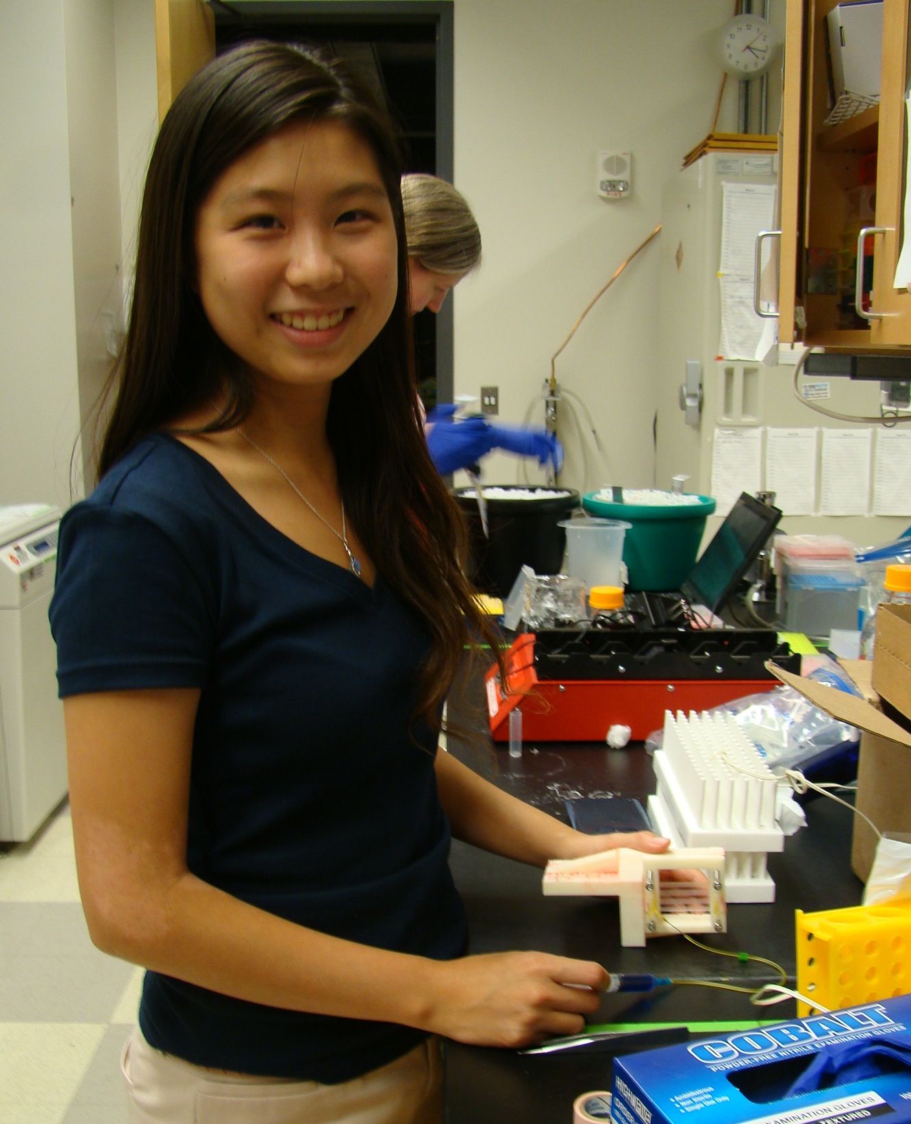
Project Title: Flow Cell and Sample Stage Design for Dual Beam Optical Tweezers for Single Molecule Experiment
Abstract: Optical tweezers are applications of physics to study the movement of biological molecules, such as DNA strands to better understand their mechanical properties. This paper aims to feature the components and apparatus set up for the optical tweezers with special emphasis on three-channel sample chamber and trapping apparatus. The optical force from the momentum of light photons emitted by the laser traps two polystyrene beads which then tethers the DNA strand and is imaged by a CCD camera.
Name: Katherine Chien
Undergraduate Institution: University of Western Ontario
Major: Medical Science with minor in Business Administration
REU Advisor: Dr. Michael Poirier
Project Title: Flow Cell and Sample Stage Design for Dual Beam Optical Tweezers for Single Molecule Experiment
Abstract: Optical tweezers are applications of physics to study the movement of biological molecules, such as DNA strands to better understand their mechanical properties. This paper aims to feature the components and apparatus set up for the optical tweezers with special emphasis on three-channel sample chamber and trapping apparatus. The optical force from the momentum of light photons emitted by the laser traps two polystyrene beads which then tethers the DNA strand and is imaged by a CCD camera.
Name: Geoffrey Green
Undergraduate Institution: Ohio State University
Major: Electrical and Computer Engeneering
REU Advisor: Dr. Roland Kawakami
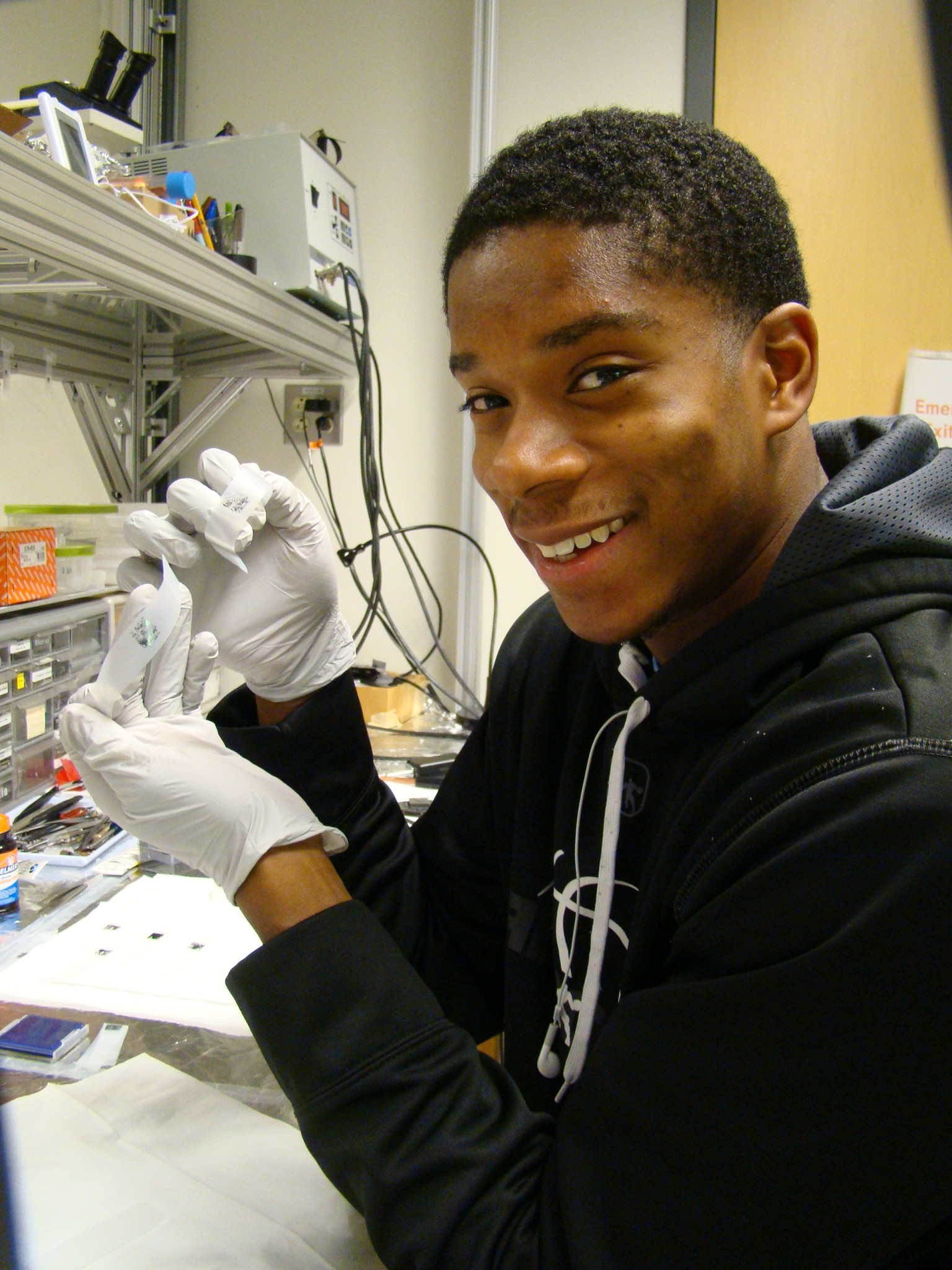
Project Title: Optimization of the Laser Writer for Nanoscale Devices
Abstract: The main goal was to compare the capabilities of the laser writer with the e-beam in terms of device fabrication. The laser writer performed best when the bake time, laser exposure, and develop time was altered, producing a sharper pattern; however, not as precise as the e-beam. Further tests will be conducted on the electrical measurements to compare the performance of devices made via e-beam and laser writer.
 Name: Toney Hicks
Undergraduate Institution: University of Alabama at Birmingham
Major: Electrical Engineering
REU Advisor: Dr. Pat Woodward
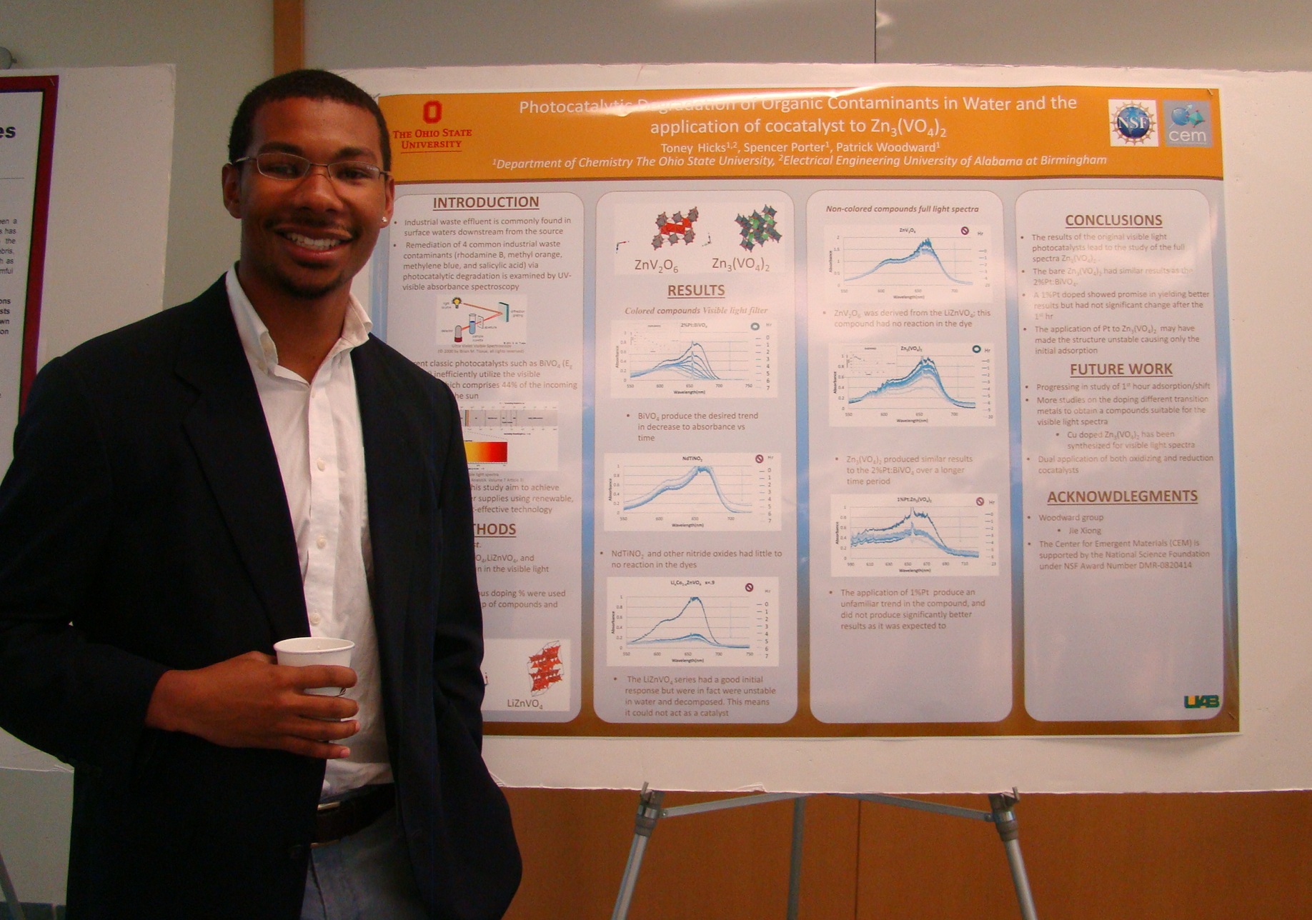
Project Title: Photocatalytic Degradation of Organic Contaminants in Water and the application of co-catalyst to Zn3(VO4)2
Abstract: Industrial waste effluent is commonly found in surface waters downstream from the source. Remediation of 4 common industrial waste contaminants (rhodamine B, methyl orange, methylene blue, and salicylic acid) via photocatalytic degradation is examined by UV-visible absorbance spectroscopy and luminescence spectroscopy. The decontamination of water is driven photocatalytically through redox reactions on the catalyst surface. Current classic photocatalysts such as BiVO4 (Eg = 2.4 eV) inefficiently utilize the visible spectrum, which comprises 44% of the incoming photons from the sun. To enhance solar light adsorption colored vanadate compounds are investigated for their activity towards degradation of organic contaminants. To further probe and enhance efficiency, platinum and other various cocatalysts are photodeposited. The application of photodeposited cocatalyst and their effects on Zn3 (VO4)2 were the final focus of the study. The compounds in this study aim to achieve better recycled water supplies using renewable, sustainable, and cost-effective technology.
Name: Toney Hicks
Undergraduate Institution: University of Alabama at Birmingham
Major: Electrical Engineering
REU Advisor: Dr. Pat Woodward
Project Title: Photocatalytic Degradation of Organic Contaminants in Water and the application of co-catalyst to Zn3(VO4)2
Abstract: Industrial waste effluent is commonly found in surface waters downstream from the source. Remediation of 4 common industrial waste contaminants (rhodamine B, methyl orange, methylene blue, and salicylic acid) via photocatalytic degradation is examined by UV-visible absorbance spectroscopy and luminescence spectroscopy. The decontamination of water is driven photocatalytically through redox reactions on the catalyst surface. Current classic photocatalysts such as BiVO4 (Eg = 2.4 eV) inefficiently utilize the visible spectrum, which comprises 44% of the incoming photons from the sun. To enhance solar light adsorption colored vanadate compounds are investigated for their activity towards degradation of organic contaminants. To further probe and enhance efficiency, platinum and other various cocatalysts are photodeposited. The application of photodeposited cocatalyst and their effects on Zn3 (VO4)2 were the final focus of the study. The compounds in this study aim to achieve better recycled water supplies using renewable, sustainable, and cost-effective technology.
 Name: Alexandra LeViness
Undergraduate Institution: University of Alabama
Major: Physics
REU Advisor: Dr. David McComb
Project Title: Processing of FIB/SEM Image Stacks from Biological Samples
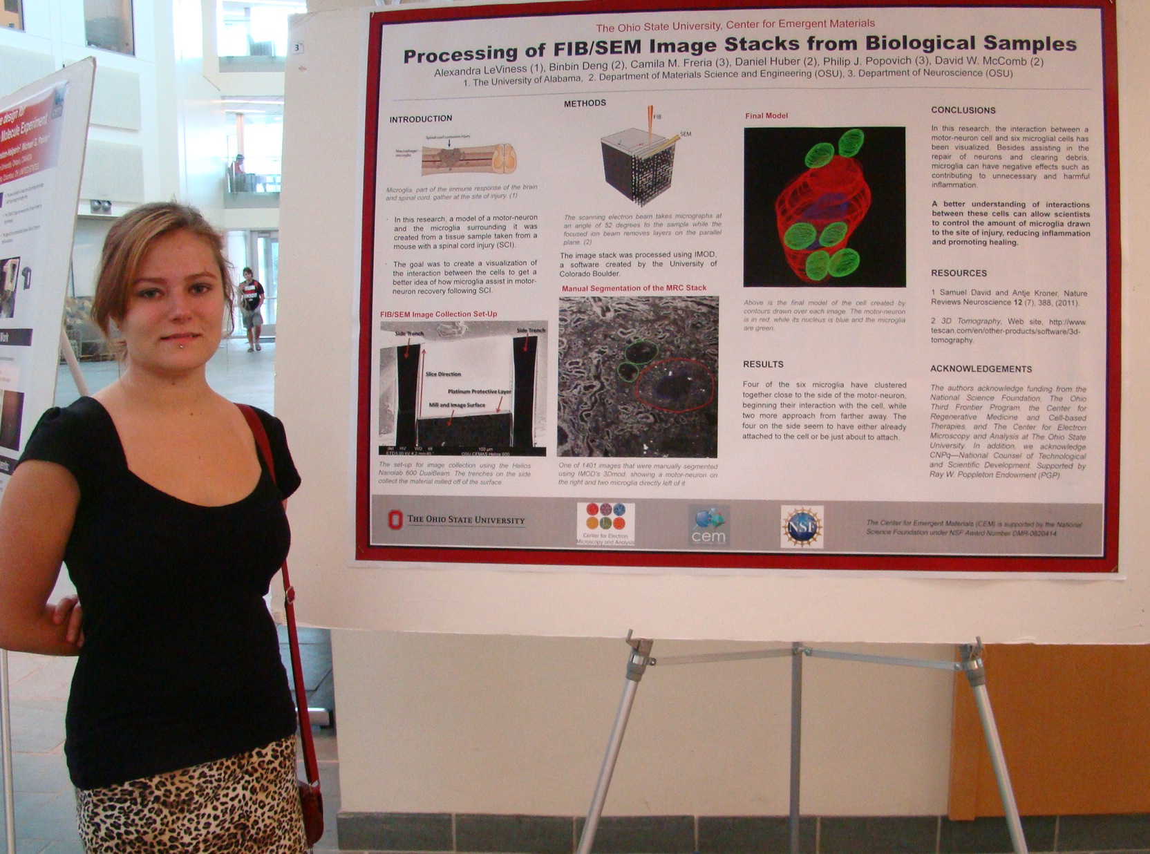
Abstract: Because of the spinal cord’s crucial role in transferring signals from the brain to the rest of the body, spinal cord injuries (SCI) are considered medical emergencies and can have effects as extreme as total paralysis below the injury1. Microglia and microphages play both beneficial and detrimental roles in the recovery process, as they clear away dead cells and myelin debris to promote recovery2, but also produce an excessive inflammatory response that can contribute to cell death3. SCI in mice serve as accurate models for those in humans4, and prior research has found that inhibiting the fractalkine receptor CX3CR1 in mice can reduce inflammation at the site of injury while contributing to synapse preservation5.
However, more research must be done to determine whether CX3CR1 inhibition can truly increase significant axon regeneration and therefore promote the reformation of functional synapses. As popular techniques such as transmission electron microscopy (TEM) are limited to small sample volumes, the use of focused ion beams and scanning electron microscopy (FIB/SEM) in the analysis of biological samples is becoming more common. This method allows for accurate 3D imaging of relatively large samples6. In this specific project, image data taken from a spinal cord sample of an SCI mouse was processed, using free EM processing software such as IMOD, in order to build a 3D model of a specific motor-neuron and the six microglia surrounding it. Four are found clustered together, possibly attached to the side of the neuron, while two more approach from another direction.
Name: Alexandra LeViness
Undergraduate Institution: University of Alabama
Major: Physics
REU Advisor: Dr. David McComb
Project Title: Processing of FIB/SEM Image Stacks from Biological Samples
Abstract: Because of the spinal cord’s crucial role in transferring signals from the brain to the rest of the body, spinal cord injuries (SCI) are considered medical emergencies and can have effects as extreme as total paralysis below the injury1. Microglia and microphages play both beneficial and detrimental roles in the recovery process, as they clear away dead cells and myelin debris to promote recovery2, but also produce an excessive inflammatory response that can contribute to cell death3. SCI in mice serve as accurate models for those in humans4, and prior research has found that inhibiting the fractalkine receptor CX3CR1 in mice can reduce inflammation at the site of injury while contributing to synapse preservation5.
However, more research must be done to determine whether CX3CR1 inhibition can truly increase significant axon regeneration and therefore promote the reformation of functional synapses. As popular techniques such as transmission electron microscopy (TEM) are limited to small sample volumes, the use of focused ion beams and scanning electron microscopy (FIB/SEM) in the analysis of biological samples is becoming more common. This method allows for accurate 3D imaging of relatively large samples6. In this specific project, image data taken from a spinal cord sample of an SCI mouse was processed, using free EM processing software such as IMOD, in order to build a 3D model of a specific motor-neuron and the six microglia surrounding it. Four are found clustered together, possibly attached to the side of the neuron, while two more approach from another direction.
 Name: Jacob Repicky
Undergraduate Institution: Pennsylvania State University
Major: Physics
REU Advisor: Dr. Jay Gupta
Project Title: Toward Characterization of Novel 2D Materials by STM
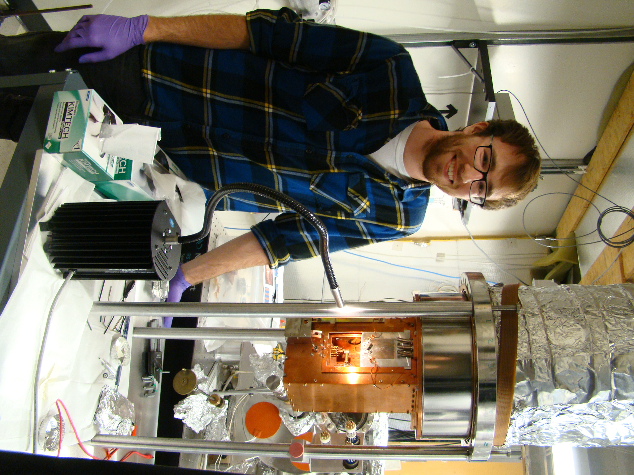
Abstract: A custom-built scanning tunneling microscope (STM) employs a piezoelectric coarse motor, for precise positioning of the probe tip near the sample surface, as well as a piezoelectric scan tube for fine, scanning motion. The scan piezo is used for acquisition and must be calibrated for proper functionality and accurate data. Topographic step height information of highly ordered pyrolytic graphite (HOPG) is gathered in air and at room temperature, then used to calibrate the motion of the scan tube along the z-axis. The xy-motion can be similarly calibrated via atomic resolution of the hexagonal graphite lattice. With a fully calibrated instrument the STM can then be used to take data on a sample of germanane grown via molecular beam epitaxy (MBE).
Name: Jacob Repicky
Undergraduate Institution: Pennsylvania State University
Major: Physics
REU Advisor: Dr. Jay Gupta
Project Title: Toward Characterization of Novel 2D Materials by STM
Abstract: A custom-built scanning tunneling microscope (STM) employs a piezoelectric coarse motor, for precise positioning of the probe tip near the sample surface, as well as a piezoelectric scan tube for fine, scanning motion. The scan piezo is used for acquisition and must be calibrated for proper functionality and accurate data. Topographic step height information of highly ordered pyrolytic graphite (HOPG) is gathered in air and at room temperature, then used to calibrate the motion of the scan tube along the z-axis. The xy-motion can be similarly calibrated via atomic resolution of the hexagonal graphite lattice. With a fully calibrated instrument the STM can then be used to take data on a sample of germanane grown via molecular beam epitaxy (MBE).
 Name: Samuel Seelbach
Undergraduate Institution: Columbus State Community College
Major: Biomedical Engineering
REU Advisor: Dr. Chris Hammel
Project Title: Development of Pulsed Techniques for Manipulation and Detection of Spin Resonance in Nitrogen-Vacancy Centers
Abstract: Nitrogen-vacancy (NV) centers in diamond are an impressive new tool that could lead to numerous scientific advancements in biological imaging using ultrasensitive magnetometry and in quantum computing. Magnetic resonance is a powerful technique that has been studied and used extensively for decades (e.g. MRI). In our experiment we exploit the dependence of NV center photoluminescence (PL) upon the spin state for optical detection of magnetic resonance from a small number of spins. We will expose NV centers to short pulses of laser and microwave radiation to manipulate the spin and then measure the overall spin state through the change in PL. Pulse techniques allow for more precise detection and direct measurement of spin lifetime than conventional electron paramagnetic resonance (EPR). Rabi and Hahn pulse protocols will be used to measure the amplitude of an internal magnetic field and the spin lifetimes. These techniques precisely manipulate spin states and overcome environmental effects. This paper discusses the progress we have made in developing an optical pulse setup for NV centers.
Name: Samuel Seelbach
Undergraduate Institution: Columbus State Community College
Major: Biomedical Engineering
REU Advisor: Dr. Chris Hammel
Project Title: Development of Pulsed Techniques for Manipulation and Detection of Spin Resonance in Nitrogen-Vacancy Centers
Abstract: Nitrogen-vacancy (NV) centers in diamond are an impressive new tool that could lead to numerous scientific advancements in biological imaging using ultrasensitive magnetometry and in quantum computing. Magnetic resonance is a powerful technique that has been studied and used extensively for decades (e.g. MRI). In our experiment we exploit the dependence of NV center photoluminescence (PL) upon the spin state for optical detection of magnetic resonance from a small number of spins. We will expose NV centers to short pulses of laser and microwave radiation to manipulate the spin and then measure the overall spin state through the change in PL. Pulse techniques allow for more precise detection and direct measurement of spin lifetime than conventional electron paramagnetic resonance (EPR). Rabi and Hahn pulse protocols will be used to measure the amplitude of an internal magnetic field and the spin lifetimes. These techniques precisely manipulate spin states and overcome environmental effects. This paper discusses the progress we have made in developing an optical pulse setup for NV centers.
 Name: Brian York
Undergraduate Institution: Clark University
Major: Physics and Mathematics
REU Advisor: Dr. Nandini Trivedi
Project Title: Topologically Protected Edge States on a Honeycomb Lattice with Spin-Orbit Coupling
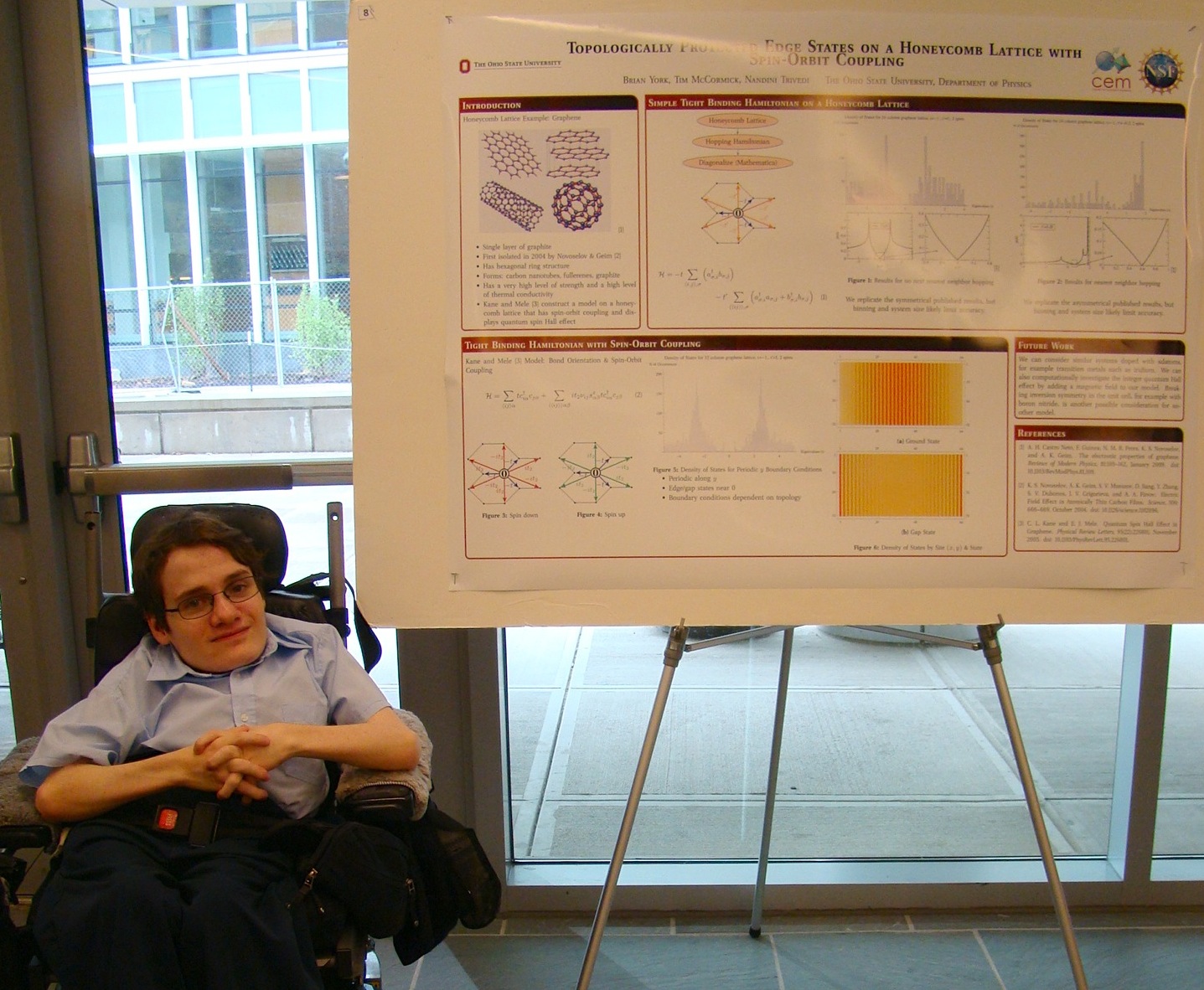
Abstract: We analyze the electronic structure of atoms on a honeycomb lattice. The kinetic energy of the system is modeled in terms of the nearest neighbor and next nearest neighbor hopping of electrons. We investigate the behavior of single-ring systems modeled as one-dimensional systems with periodic boundary conditions. This is extended to multi-ring systems and a bulk lattice. After implementing a simple model[1] neglecting spin and spin-orbit coupling and replicating the predicted band structure, we implement the Kane and Mele [2] model. This is a next nearest neighbor tight binding graphene model including next nearest neighbor hopping energies dependent on bond orientation and spin. We replicate the predicted band structure for this model as well and demonstrate that spin-orbit coupling on a honeycomb lattice with edges causes new energy states to appear in the spectrum of its Hamiltonian. These states are found at the edges of the lattice and are robust against disorder.
Name: Brian York
Undergraduate Institution: Clark University
Major: Physics and Mathematics
REU Advisor: Dr. Nandini Trivedi
Project Title: Topologically Protected Edge States on a Honeycomb Lattice with Spin-Orbit Coupling
Abstract: We analyze the electronic structure of atoms on a honeycomb lattice. The kinetic energy of the system is modeled in terms of the nearest neighbor and next nearest neighbor hopping of electrons. We investigate the behavior of single-ring systems modeled as one-dimensional systems with periodic boundary conditions. This is extended to multi-ring systems and a bulk lattice. After implementing a simple model[1] neglecting spin and spin-orbit coupling and replicating the predicted band structure, we implement the Kane and Mele [2] model. This is a next nearest neighbor tight binding graphene model including next nearest neighbor hopping energies dependent on bond orientation and spin. We replicate the predicted band structure for this model as well and demonstrate that spin-orbit coupling on a honeycomb lattice with edges causes new energy states to appear in the spectrum of its Hamiltonian. These states are found at the edges of the lattice and are robust against disorder.
 Name: Angelita Zacharias
Undergraduate Institution: The Ohio State University
Major: Biomedical Engineering
REU Advisor: Dr. Carlos Castro
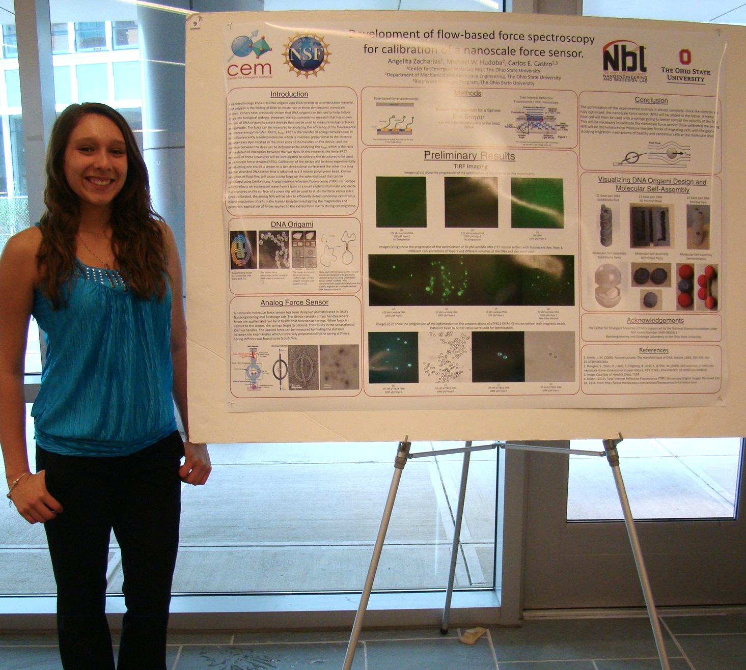
Project Title: Development of Flow‐Based Force Spectroscopy for Calibration of a Nanoscale Force Sensor
Abstract: Structural DNA nanotechnology is a rapidly emerging field that uses DNA as a construction material. DNA origami is the programmed folding of many DNA strands to create two or three dimensional, nanoscale shapes. Previous research has developed DNA origami nanostructures for a range of applications, such drug delivery. However, current applications of DNA origami focus on designing specific geometry with little consideration for mechanical function. The Nanoengineering and Biodesign Lab at Ohio State University is seeking to create devices with designed mechanical properties that can be used to measure biological forces at nanoscale. The force can be measured by analyzing the efficiency of the fluorescence resonance energy transfer (FRET), which is the distance-dependent transfer of energy between two fluorescent molecules. The goal of this research is to develop an experimental assay to calibrate the force-FRET behavior of these nanoscale force sensors (NFSs). Calibration of the NFSs requires the ability to apply a known force to the device. This will be done experimentally by attaching one end of a sensor to a two dimensional surface and the other to a long double stranded DNA tether that is attached to a ~micron size polystyrene bead. Known velocities of fluid flow will cause a drag force on the spherical bead that is transferred via the tether to the NFS. The force can be calculated using Stoke’s Law. A single molecule fluorescence microscope can be used to simultaneously monitor the FRET signal as the force is applied. Once calibrated, the NFSs can be applied to investigate molecular forces in a wide range of biological systems, for example studying the migration mechanisms of highly metastatic cancerous cells.
Name: Angelita Zacharias
Undergraduate Institution: The Ohio State University
Major: Biomedical Engineering
REU Advisor: Dr. Carlos Castro
Project Title: Development of Flow‐Based Force Spectroscopy for Calibration of a Nanoscale Force Sensor
Abstract: Structural DNA nanotechnology is a rapidly emerging field that uses DNA as a construction material. DNA origami is the programmed folding of many DNA strands to create two or three dimensional, nanoscale shapes. Previous research has developed DNA origami nanostructures for a range of applications, such drug delivery. However, current applications of DNA origami focus on designing specific geometry with little consideration for mechanical function. The Nanoengineering and Biodesign Lab at Ohio State University is seeking to create devices with designed mechanical properties that can be used to measure biological forces at nanoscale. The force can be measured by analyzing the efficiency of the fluorescence resonance energy transfer (FRET), which is the distance-dependent transfer of energy between two fluorescent molecules. The goal of this research is to develop an experimental assay to calibrate the force-FRET behavior of these nanoscale force sensors (NFSs). Calibration of the NFSs requires the ability to apply a known force to the device. This will be done experimentally by attaching one end of a sensor to a two dimensional surface and the other to a long double stranded DNA tether that is attached to a ~micron size polystyrene bead. Known velocities of fluid flow will cause a drag force on the spherical bead that is transferred via the tether to the NFS. The force can be calculated using Stoke’s Law. A single molecule fluorescence microscope can be used to simultaneously monitor the FRET signal as the force is applied. Once calibrated, the NFSs can be applied to investigate molecular forces in a wide range of biological systems, for example studying the migration mechanisms of highly metastatic cancerous cells.